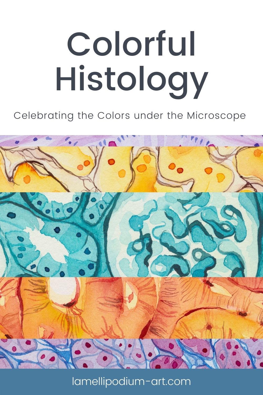
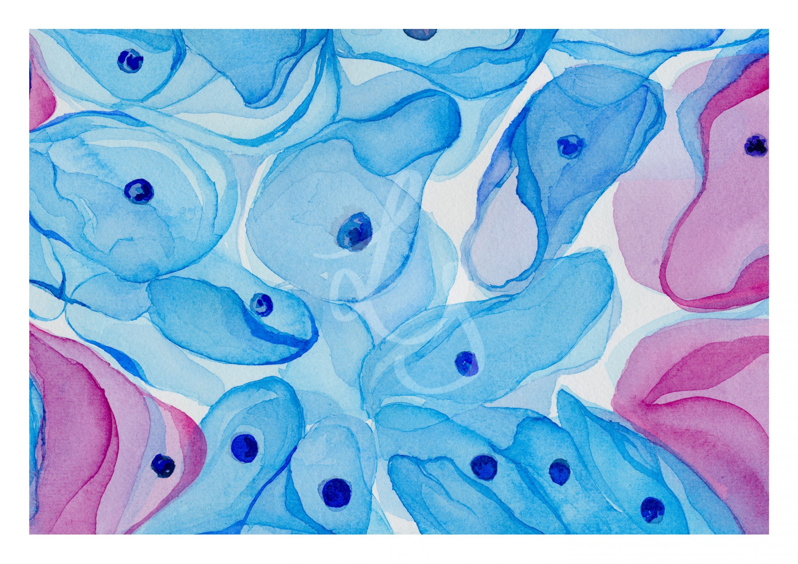
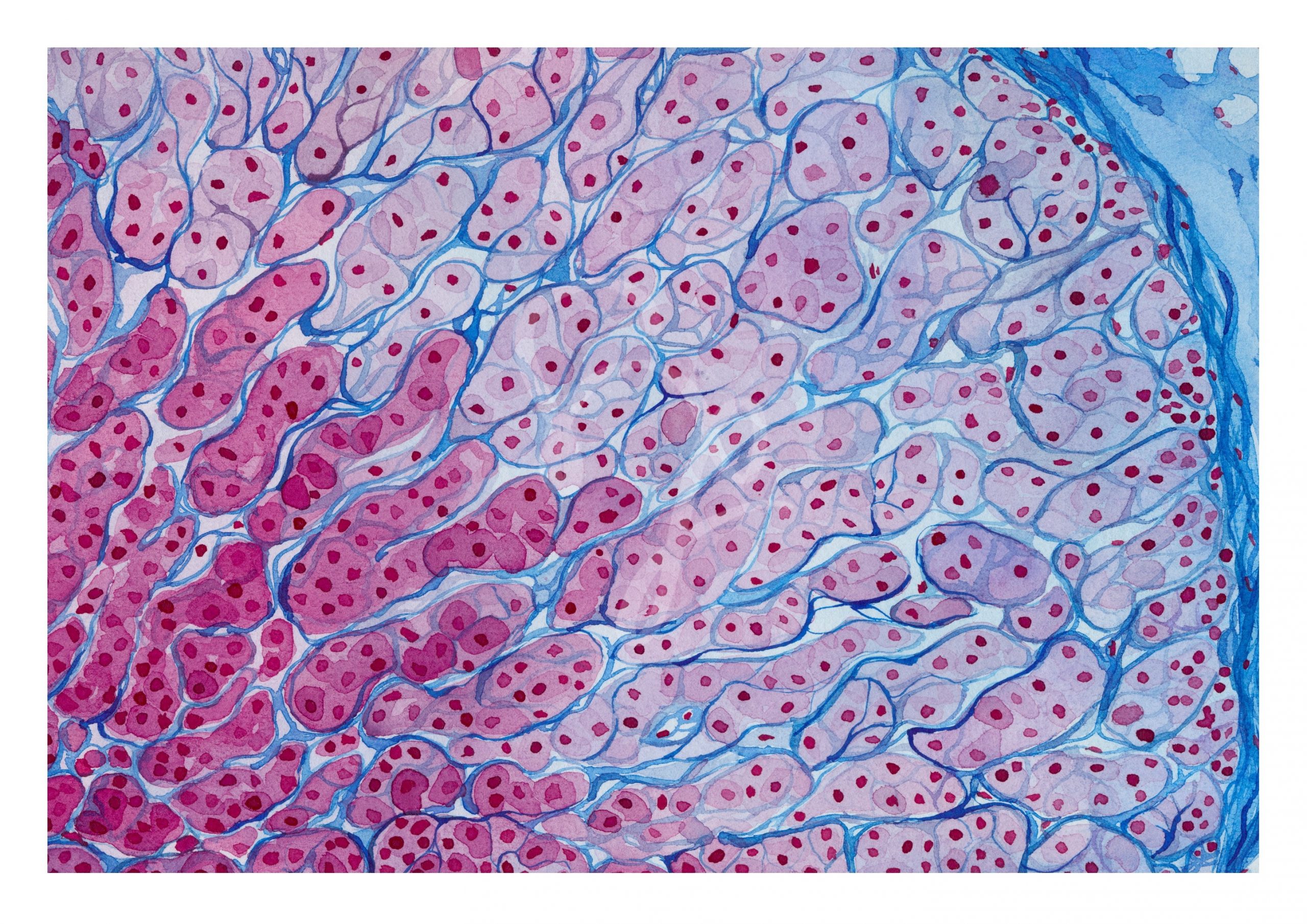
In January and February of 2021 I created a painting series around the colorful world under the microscope brought to us by all the beautiful staining methods. The series comprises nine watercolor paintings showing different nine tissues and stains.
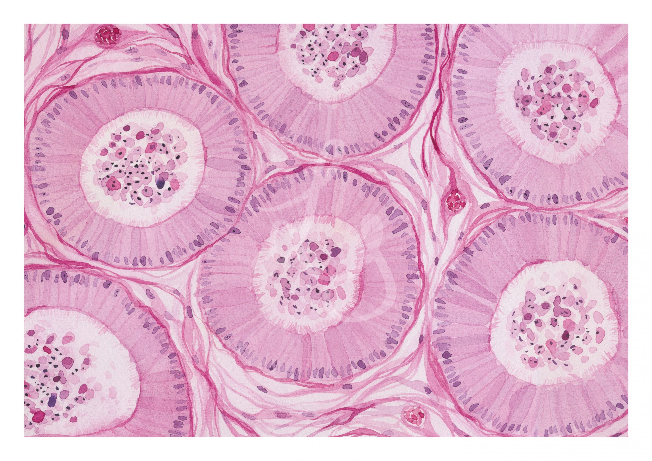
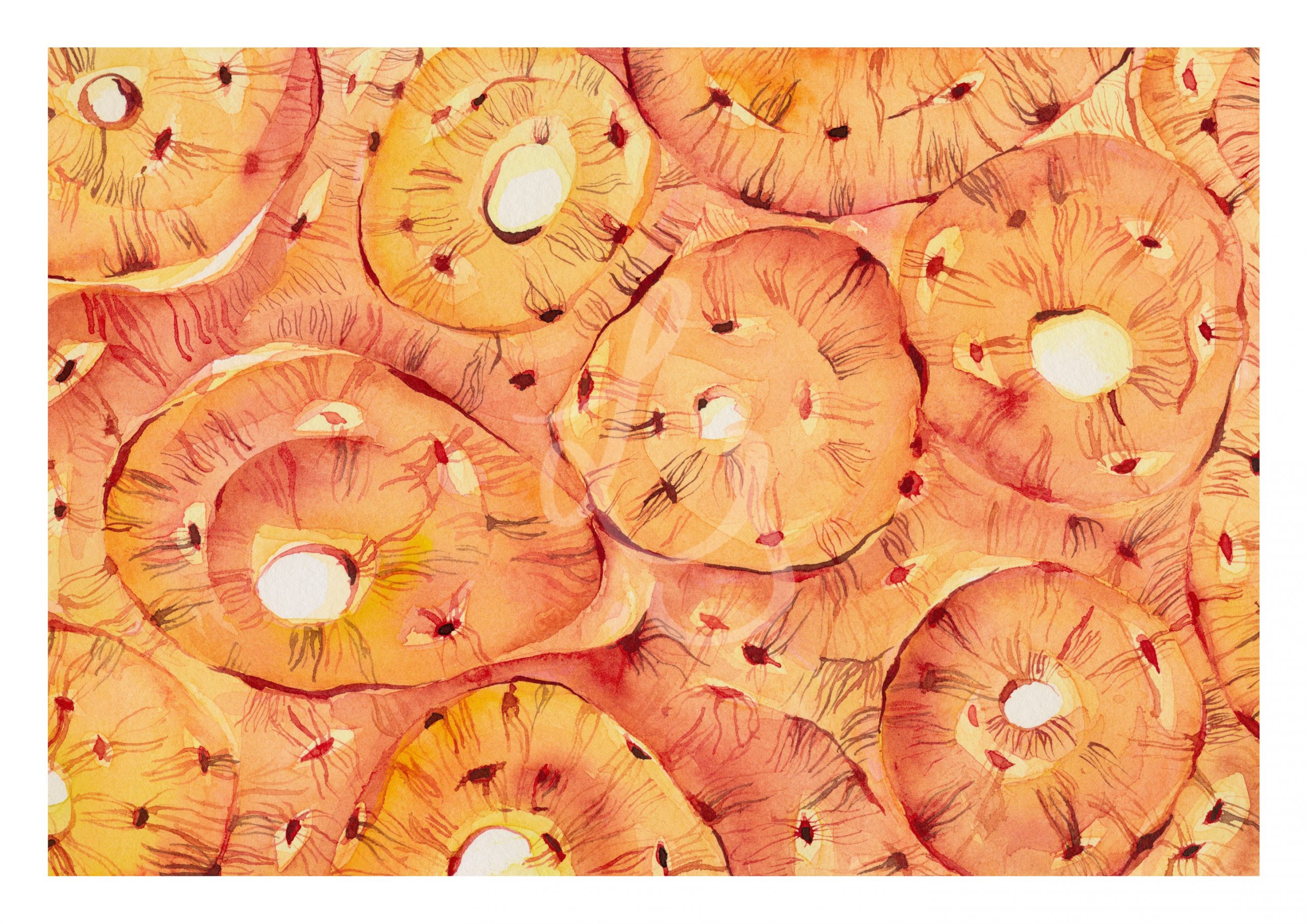
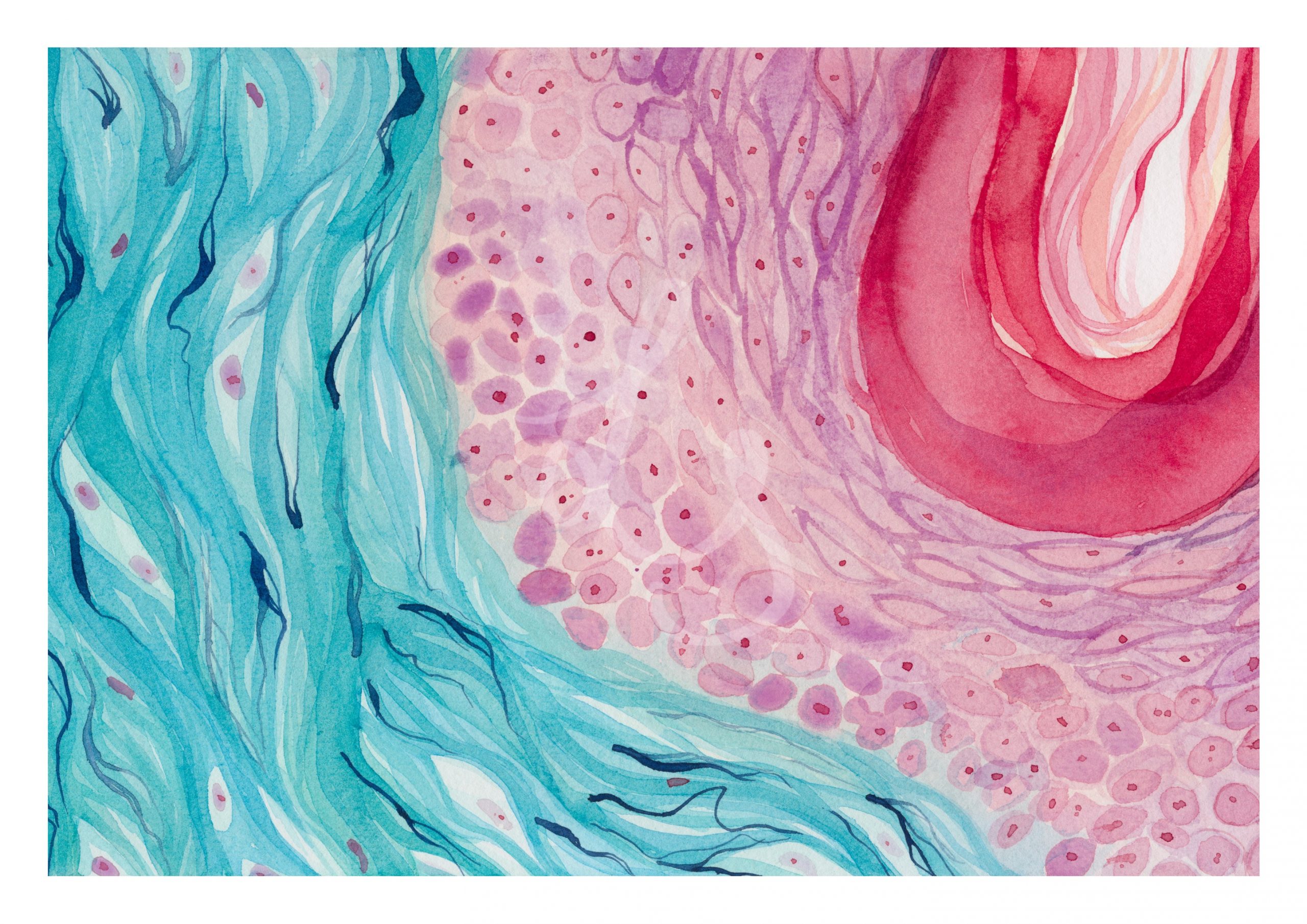
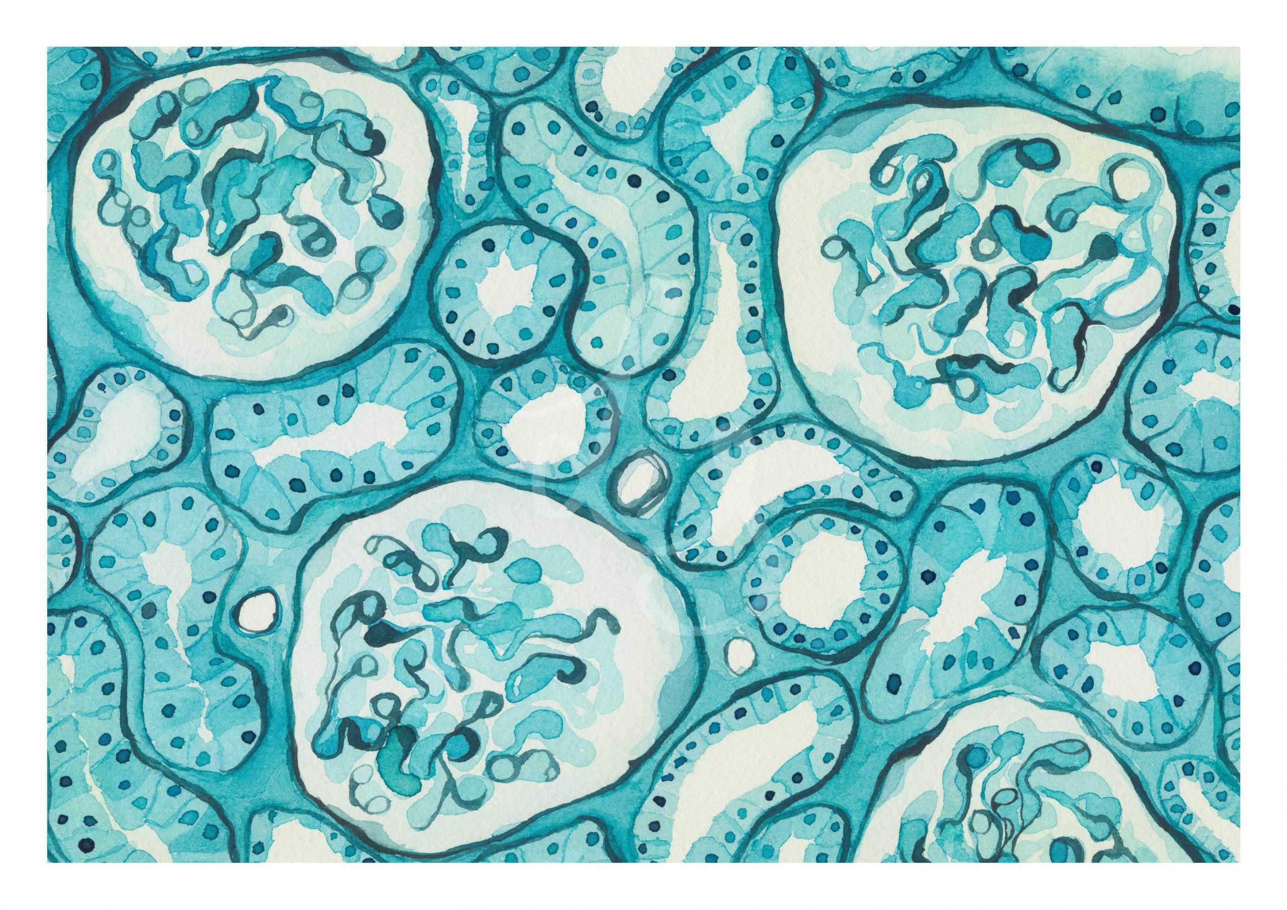
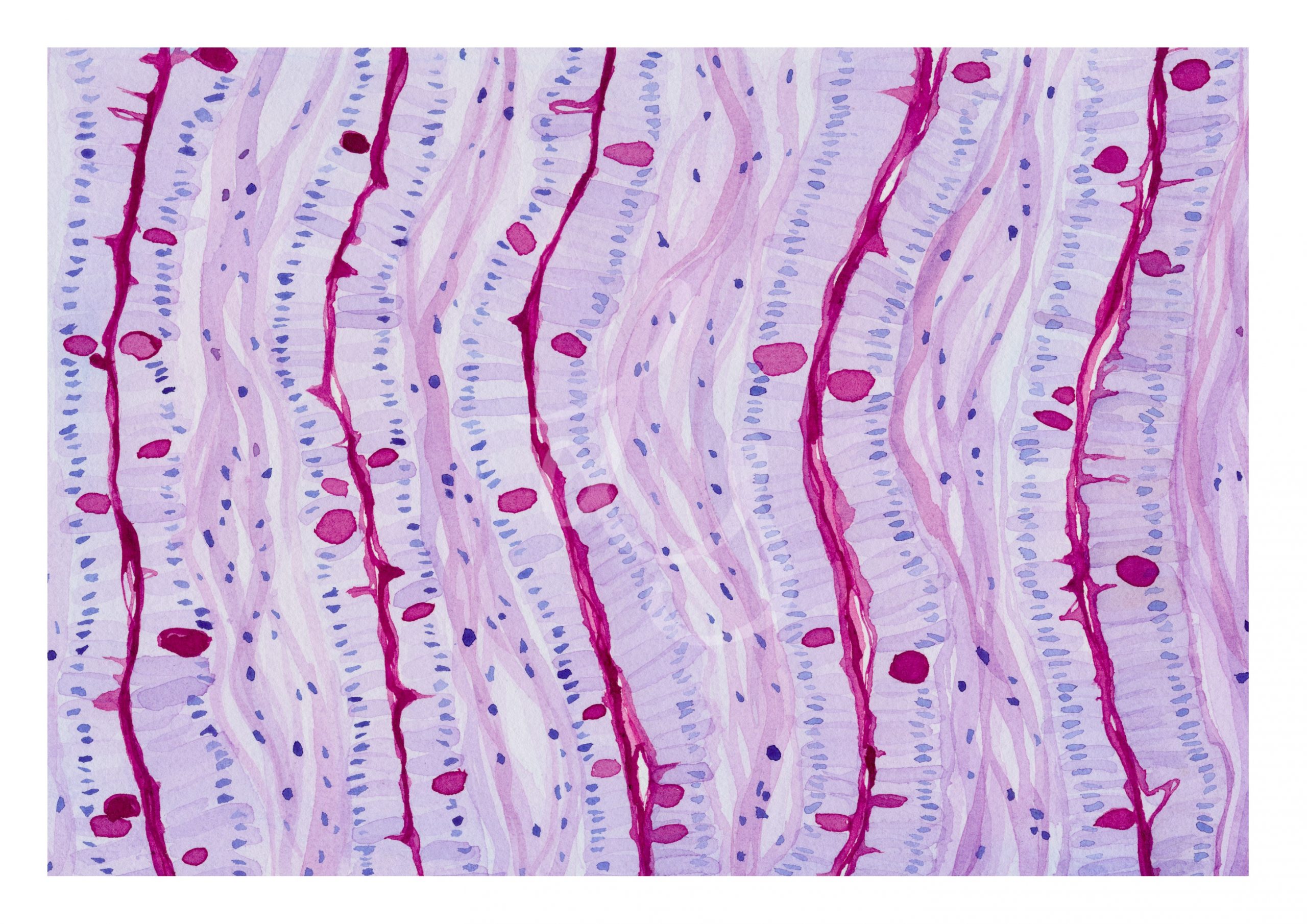
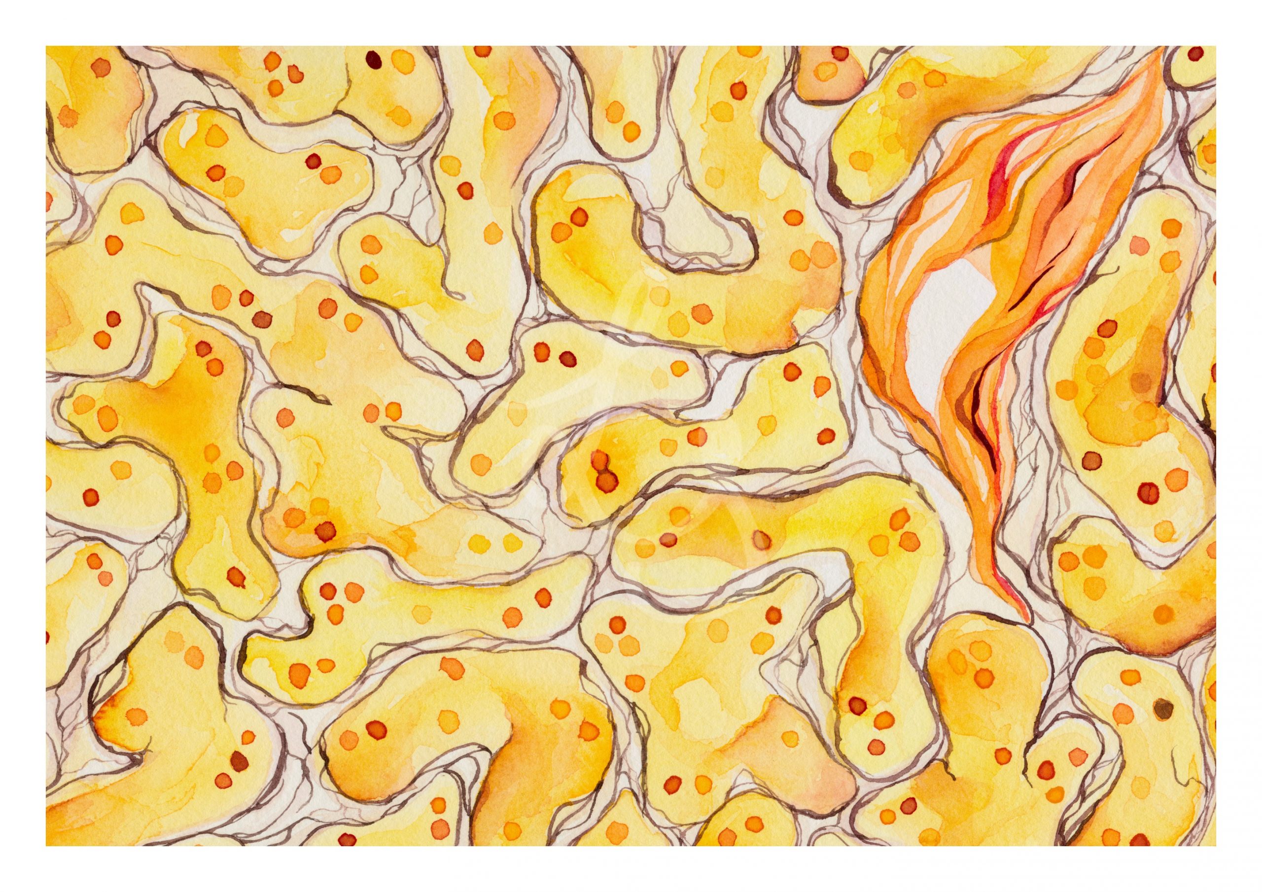
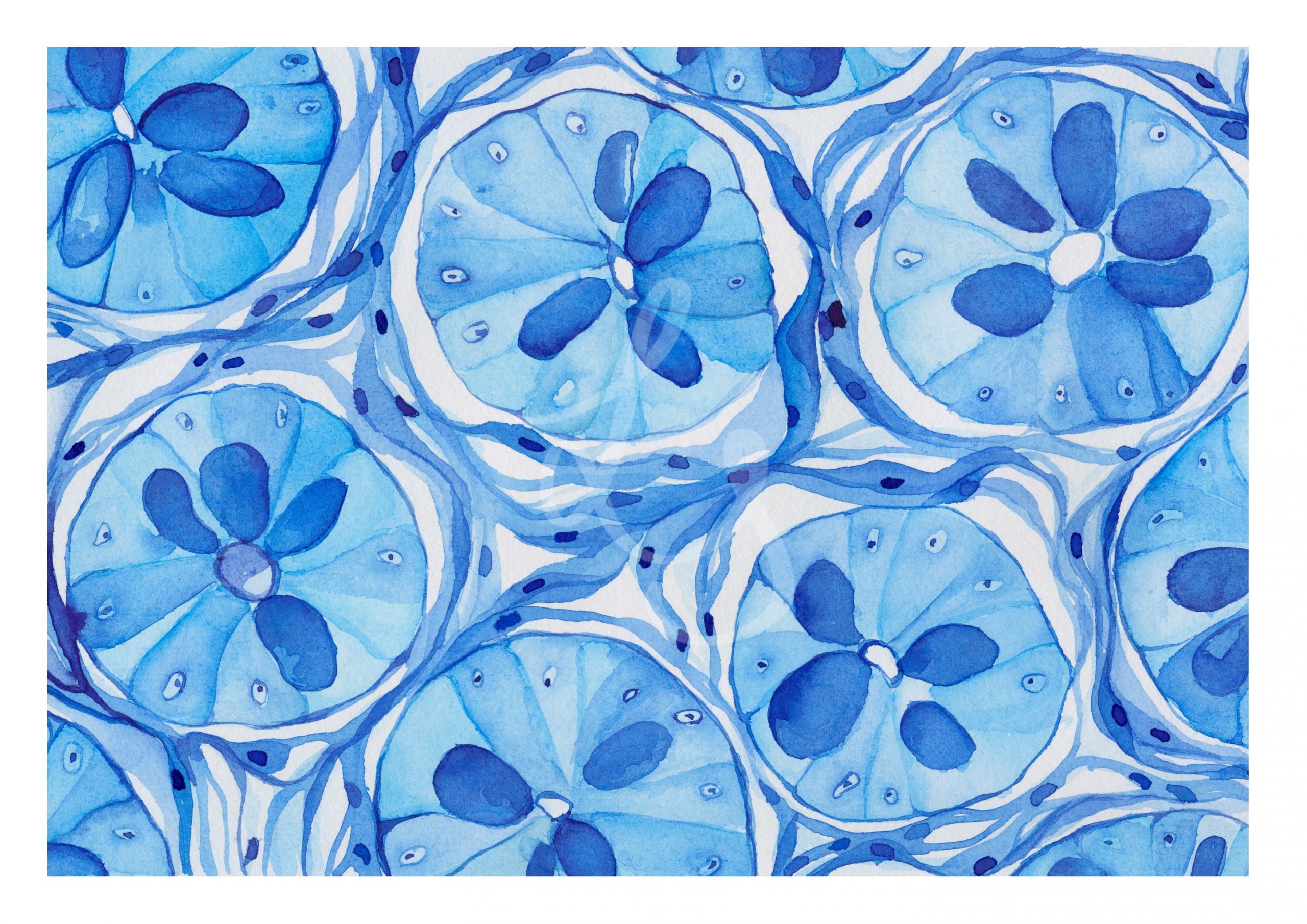
Recreating a series
If you have been following my artwork for a while you might be familiar with the first two series featuring different staining methods. The different colors and contrasts that are created by the stains are very inspiring to me and I felt an inner urge to repaint parts of this series since last autumn. I learned a lot since then and I feel like my style has not really changed much but gained in confidence and contrast. I wanted to challenge that feeling by recreating the series on a larger format and really celebrate the colorful world of histology.
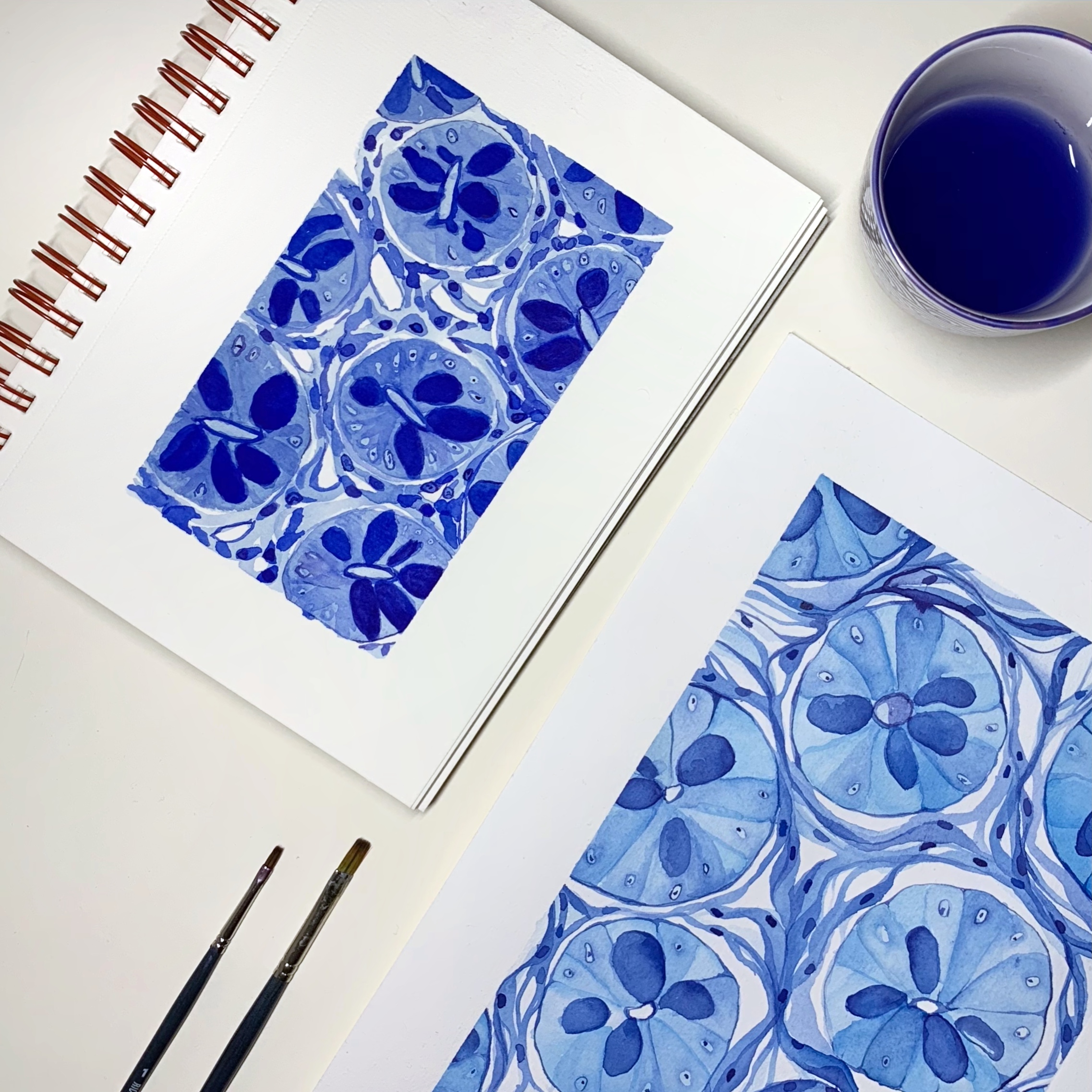
Two versions of colon stained with methylene blue: on the top, the first smaller version – on the bottom, the new, larger version.
How I chose these stains
It was not easy to pick the fitting stains and there still some left I had on the list. But I tried to pick those which go well with “normal” histology. Some stains are just more appealing to me in the context of a disease, for example, Prussian blue or Giemsa. The final nine picks are a mix of everyday stains like H&E, Pap, or PAS, but also some really special ones I have only seen for didactical reasons in Histology books, such as Thionin and Azan.
What about all the missing stains?
Since I left out quite a few stains I am positive there will be a next stain series, which will be more focused on stains in the context of pathologies. But first I will focus on some hematology work.
Update (August 21): Find the mentioned pathology series here and the mentioned blood cell paintings here!
