There are many bodies in pathology, no not only those in autopsy, I am talking about those under the microscope. Several Structures we can see in diseases have been given a „XY-body“ name, often named after the person who described them first. For this Advent Calendar Series I have chosen to paint 12 of these bodies and share them while counting down to Christmas.
1st December
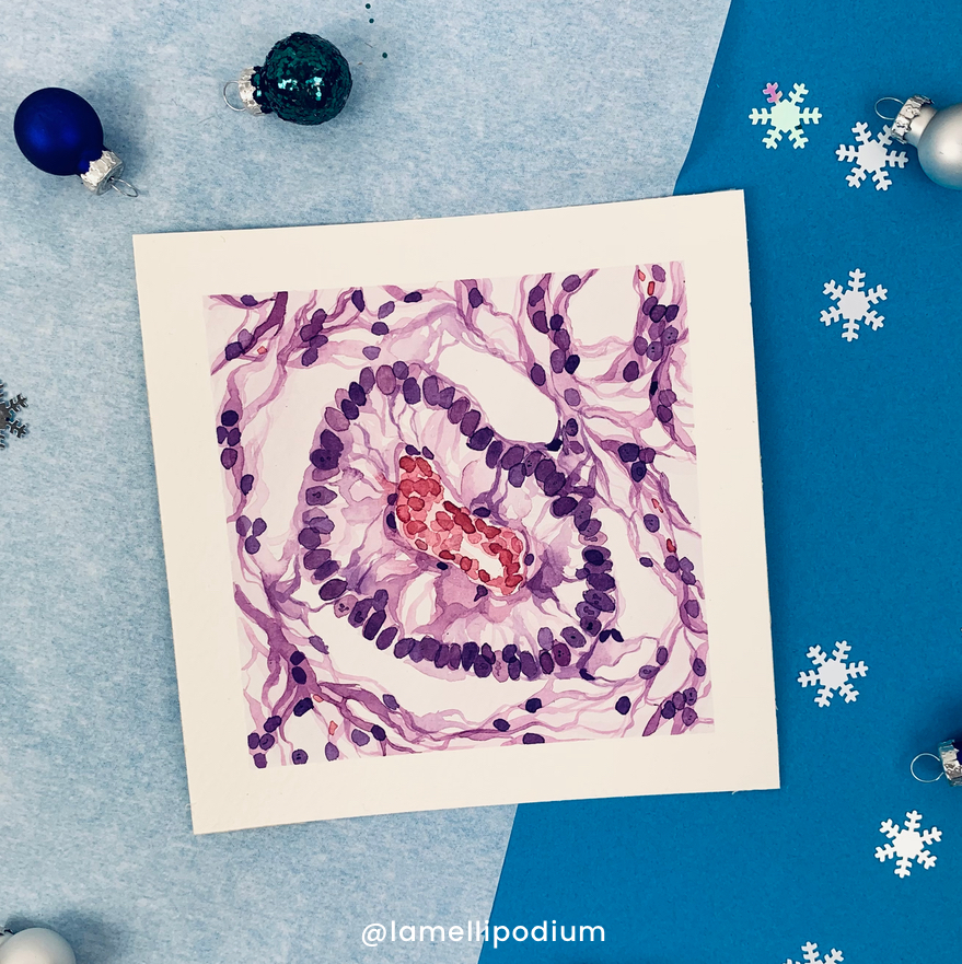
Schiller-Duval Bodies are structures that are pathognomonic for Yolk Sac tumors, also called Endodermal Sinus tumors, which are a type of Germ Cell Tumor. They have been firstly described in the late nineteenth century by Mathias-Marie Duval and Walter Schiller.
3rd December
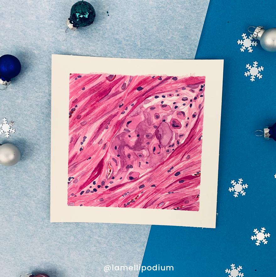
Aschoff bodies are characteristic of rheumatic heart disease. They are a special kind of granuloma found in patients with acute thematic fever. They were described independently by Ludwig Aschoff and Paul Rudolf Geipel, and for this reason, they are occasionally called Aschoff-Geipel bodies.
5th December
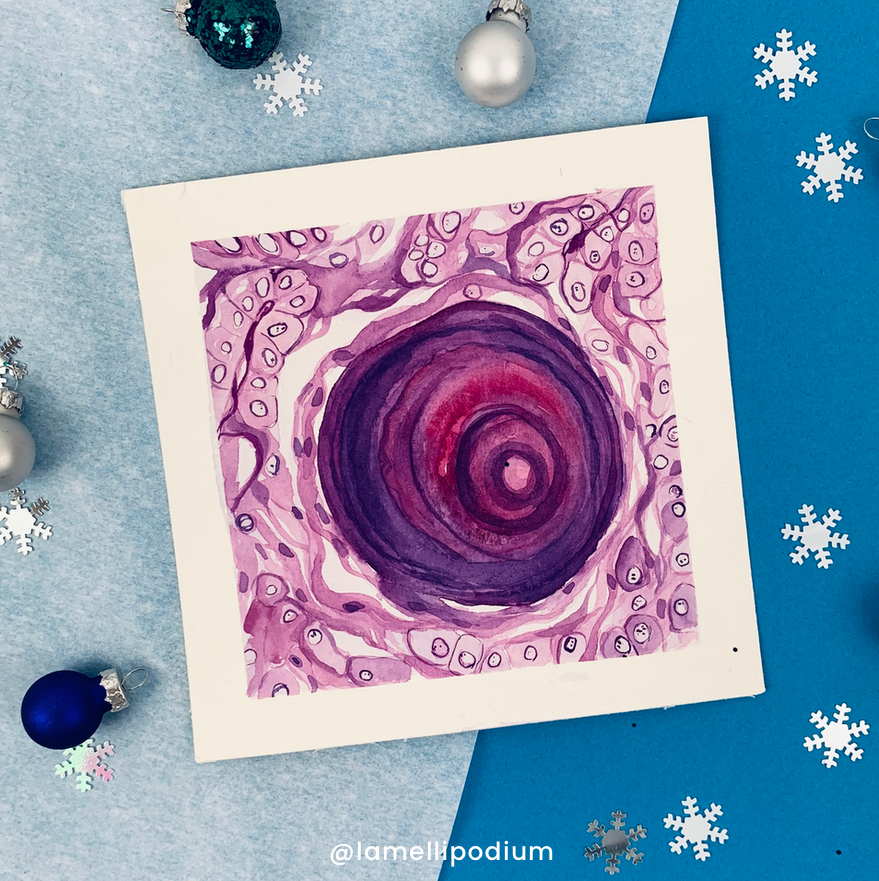
When I asked my followers on social media to name their favourite bodies, the psammoma bodies were very popular. The term psammoma is derived from the Greek word psámmos, meaning sand. They are seen in the context of a long list of diseases, including Meningiomas, and Papillary thyroid carcinoma, which I painted here.
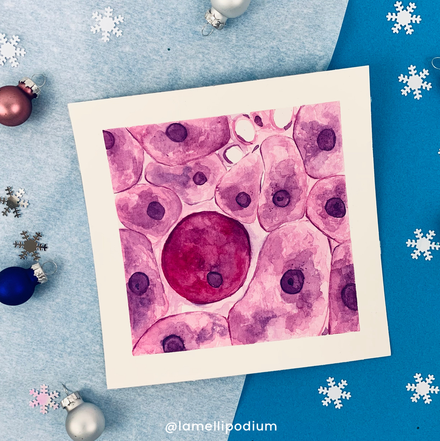
7th December
Councilman bodies are dying hepatocytes observable in patients suffering from acute viral hepatitis and other conditions. They are named after the American pathologist William Thomas Councilman who did a lot of research on yellow fever
9th December
Civatte bodies are eosinophilic, apoptotic keratinocytes in the lower epidermis and at the dermo-epidermal junction which can be found in lichen planus or other interface dermatoses. I read in a paper that they were first described by Raymond Sabouraud and for a time they were apparently called ‘Sabouraud cells’.
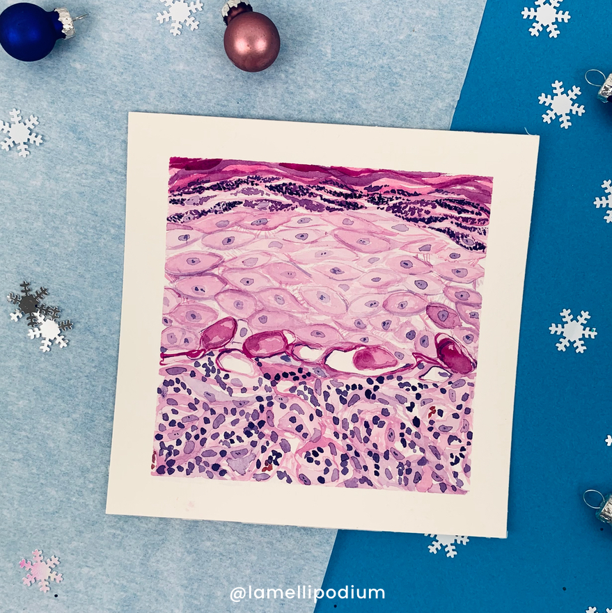
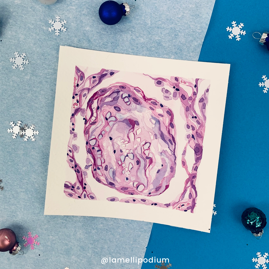
11th December
The Masson bodies are the histomorphologic hallmark of organizing pneumonia. They are polypoid plugs of loose organizing connective tissue that obliterate the airway. I can’t help myself but I like the light blue of the myxomatous connective tissue.
13th December
Mallory-Denk bodies are inclusions found in the cytoplasm of liver cells. They consist of damaged intermediate filaments and can be found in patients suffering from alcohol-induced liver disease but also in Wilsons disease or Primary biliary cholangitis.
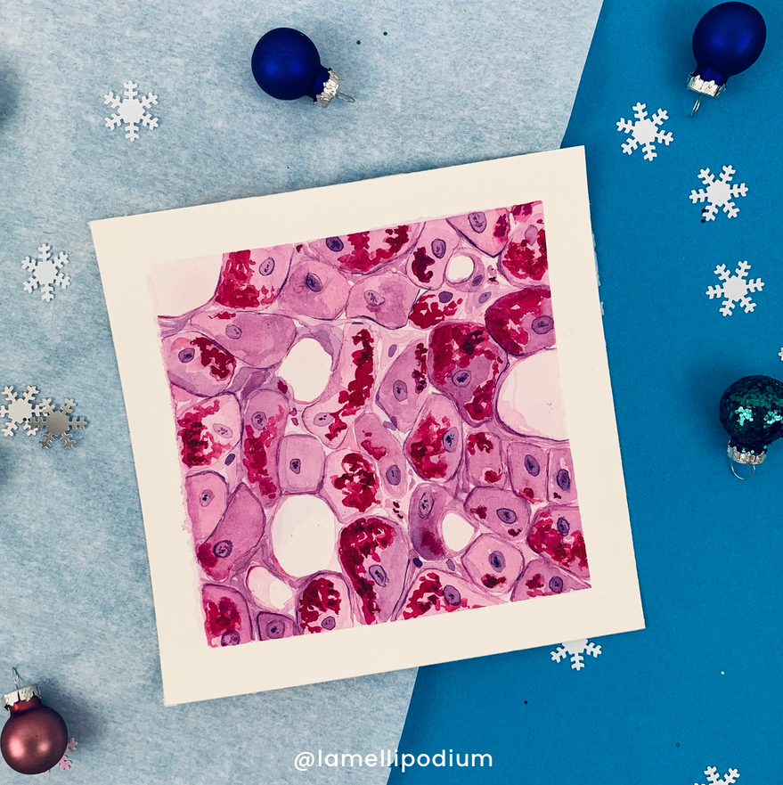
15th December
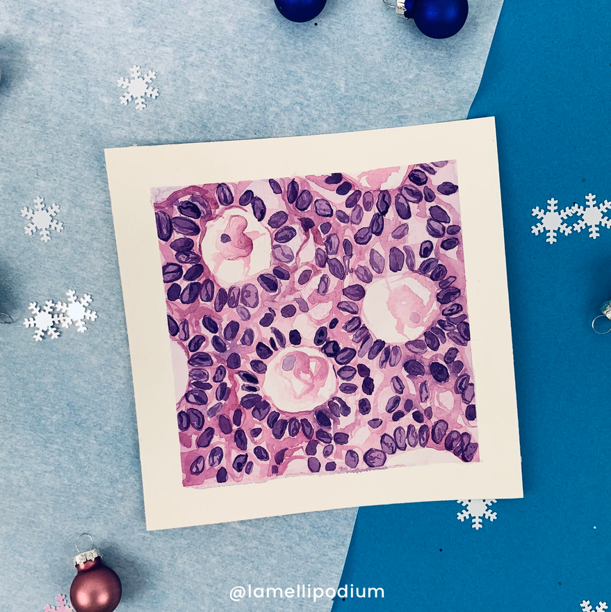
Call-Exner bodies are are small eosinophilic fluid-filled spaces between granulosa cells with carved „coffee-bean” nuclei. They are pathognomonic for granulosa cell tumors and named after Emma Louise Call and Sigmund Exner.
17th December
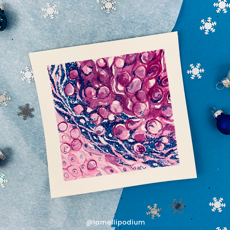
My favorites of the series: Henderson-Patterson bodies as seen in Molluscum Contagiosum. These refer to intracytoplasmic eosinophilic inclusions in keratinocytes of the stratum spinosum and granulosum. These skin lesions are often observed in young children and are caused by the molluscum contagiosum virus (MCV).
19th December
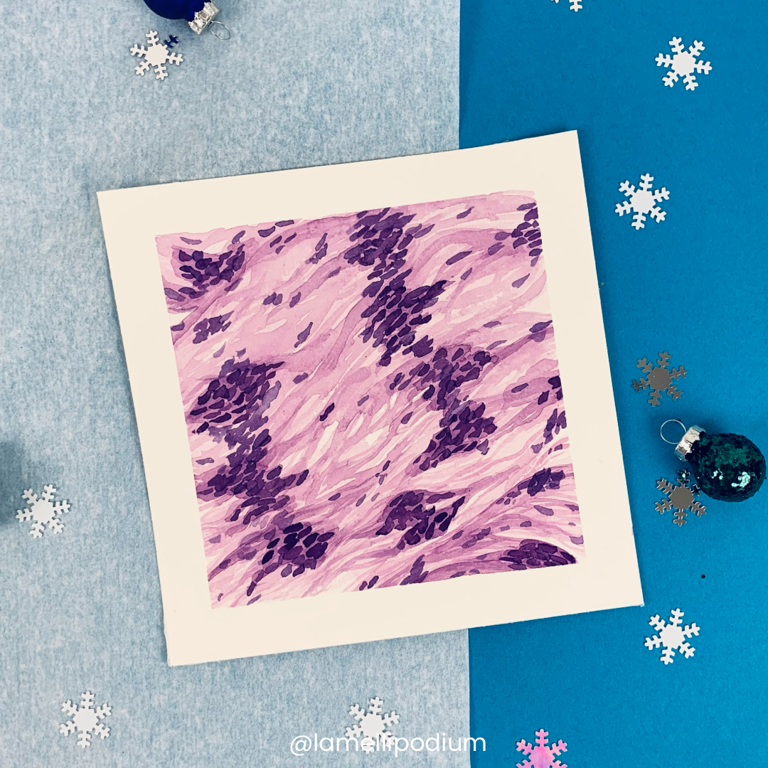
Verocay bodies are a histopathological finding in schwannomas, usually benign
nerve sheath tumors. They wer first described by Uruguayan neuro-pathologist José Juan Verocay in 1910. They are a component of the „Antoni A” areas , which refer to the dense areas of schwannomas.
21st December
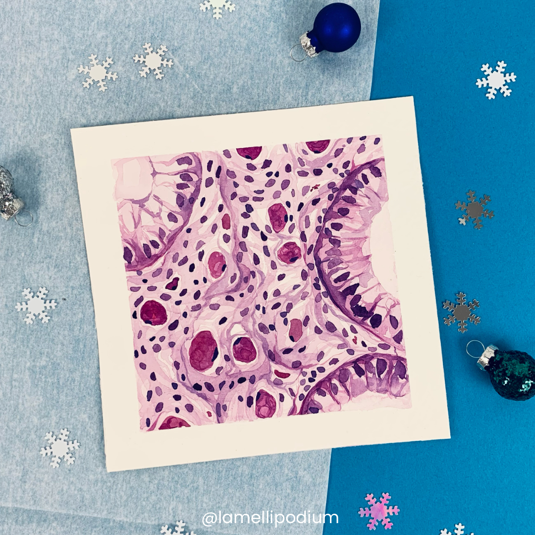
Russell bodies are inclusion bodies usually found in atypical plasma cells that become known as Mott cells. They are eosinophilic, homogeneous immunoglobulin-containing inclusions usually found in cells undergoing excessive synthesis of immunoglobulins.
23rd December
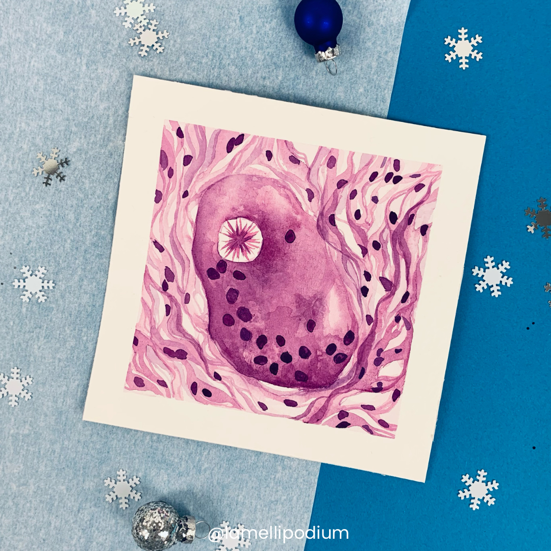
I saved the asteroid bodies for last because to me they look like little stars and what is more fitting so close to Christmas. They can be observed in giant cells in granulomatous diseases like sarcoidosis or in foreign body reactions.
