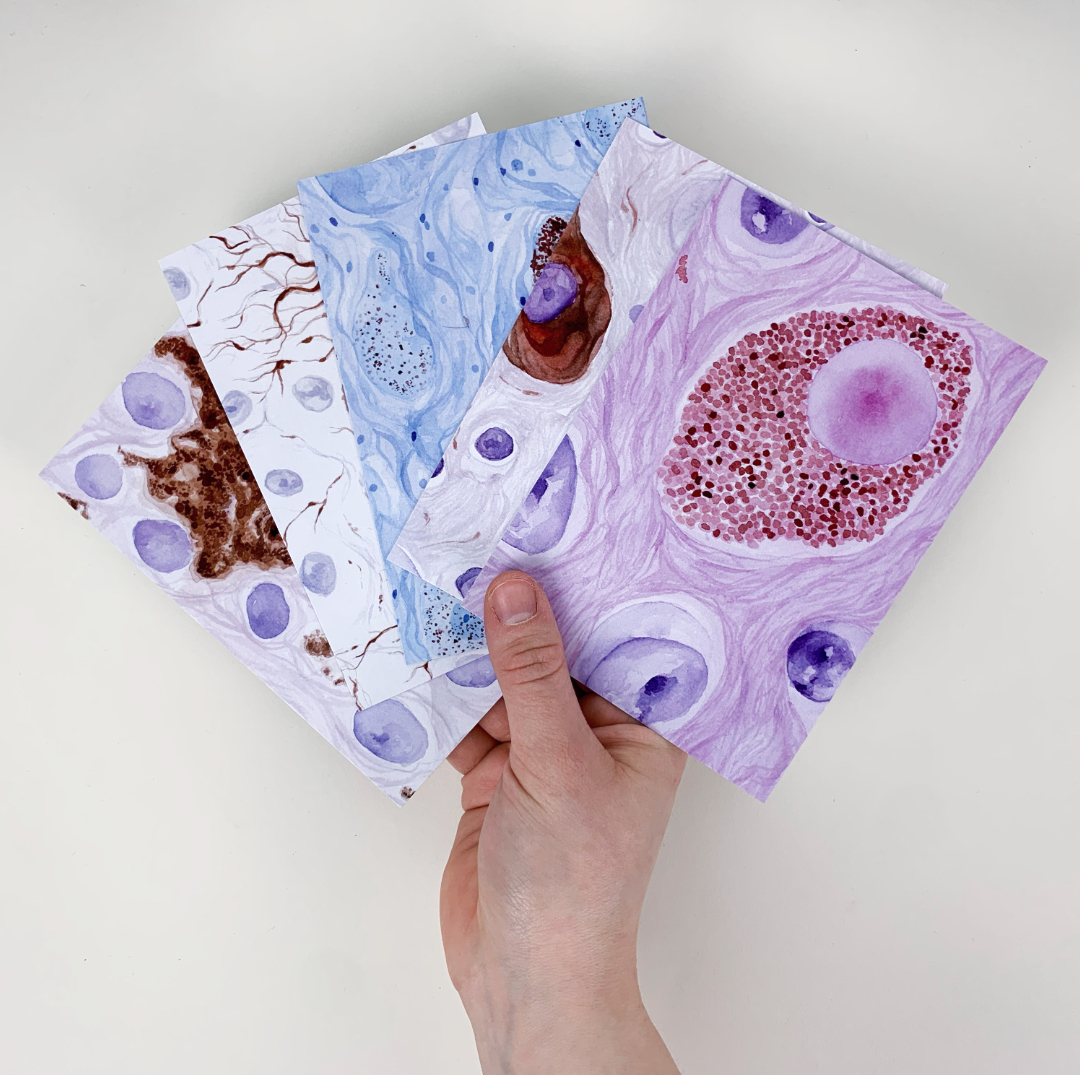
This is my second series of the year and also my second collaboration series! For this series I teamed up with Kimberly from @thepathphd. Kimberly does her PhD in Pathology and researches the role of tau in diseases. She had reached out to me about a collaboration and we decided to do a series about Neuropathology!
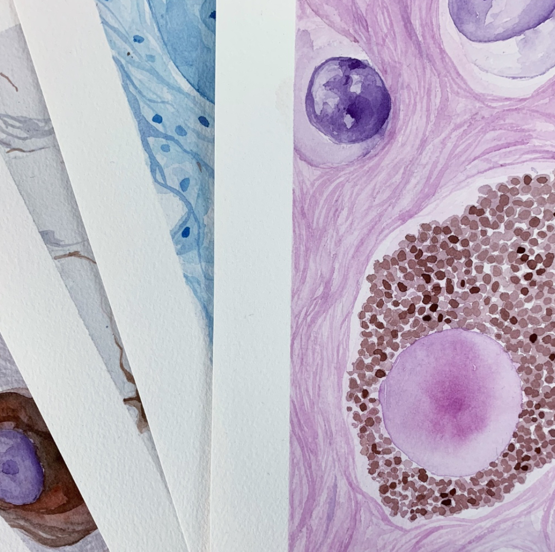
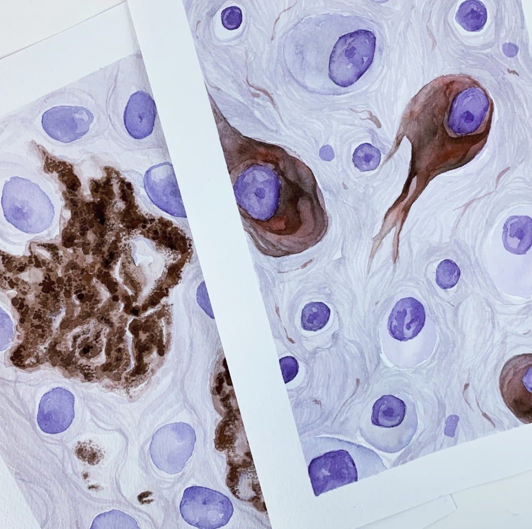
I am really happy I got the chance to work together with Kimberly as I have not had a lot of exposure to Neuropathology as it is not part of my residency. My approach to these paintings was definitely different from painting my mostly “epithelium-based” type of paintings – it felt like working in a bit more unorganized space or let’s say limitless.
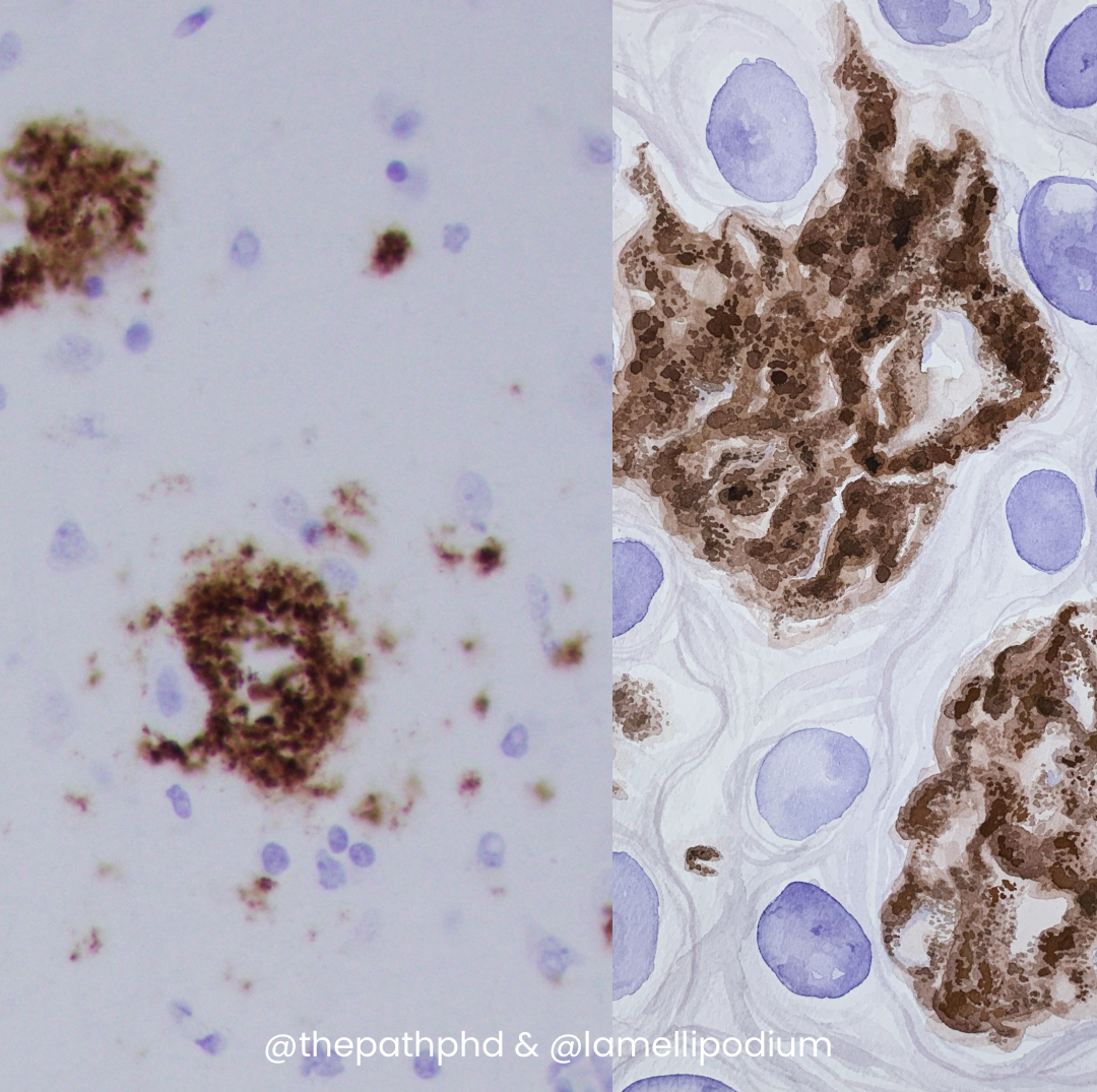
Beta-Amyloid Plaquesa
Reference on the left and painting on the right
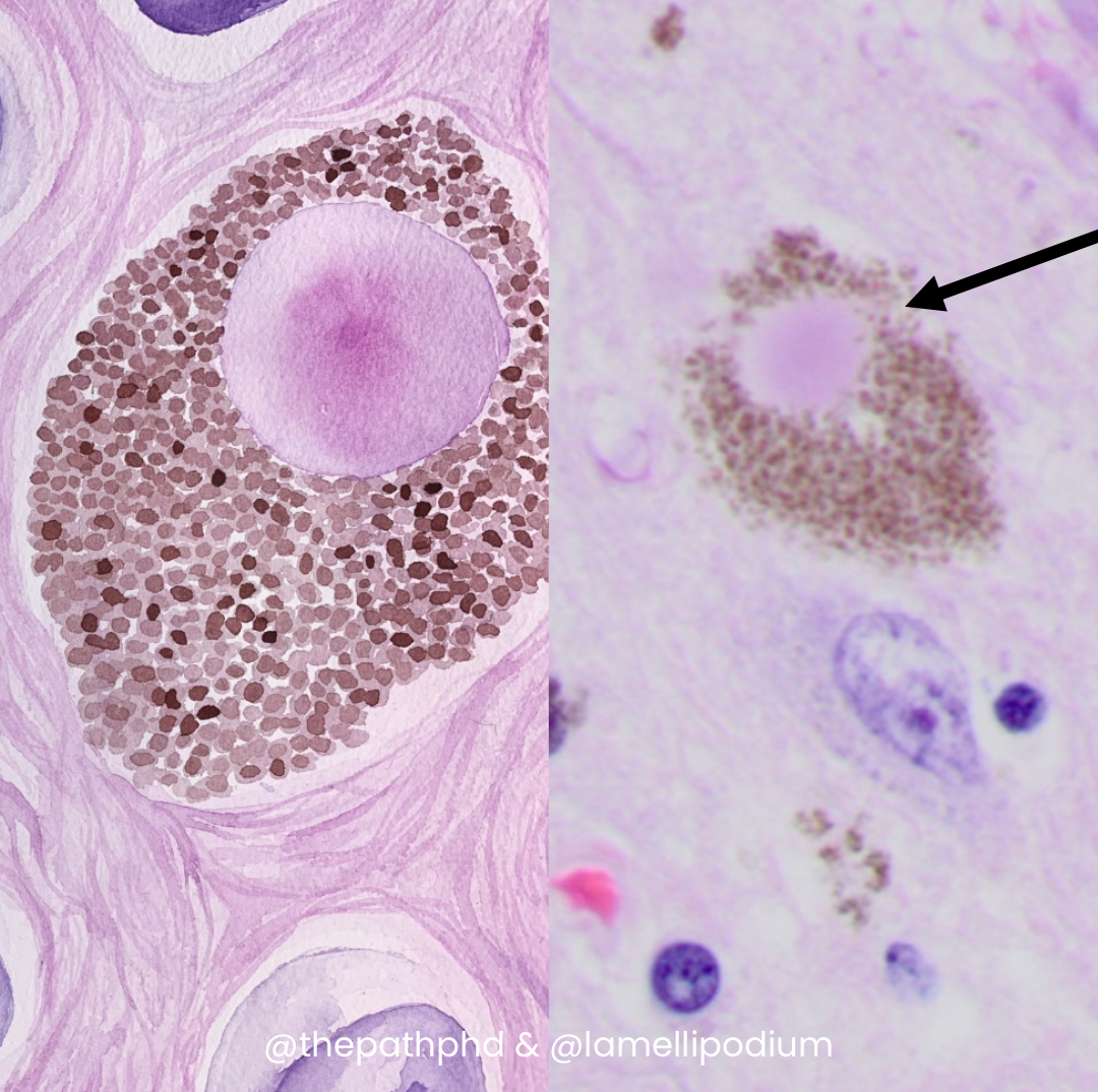
Lewy Body
Reference on the right and painting on the left
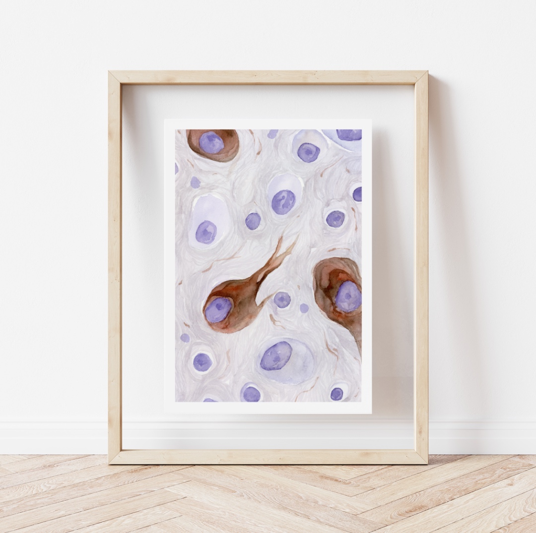
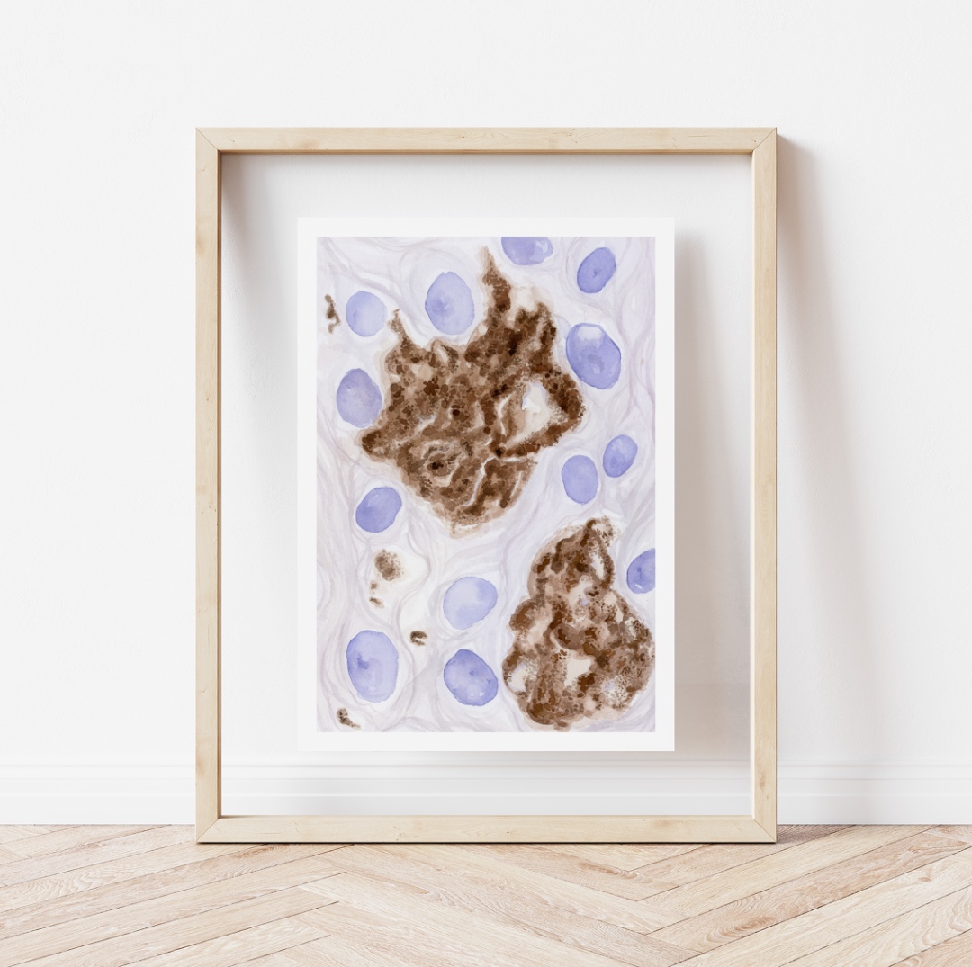
This painting was inspired by TDP-43 accumulations which can be found in a number of diseases, such as frontotemporal lobar degeneration with TDP-43 (FLTD-TDP43) and amyotrophic lateral sclerosis (ALS, pictured here). These are cytoplasmic inclusions that typically occur in the cortex, hippocampus, and amygdala in FTLD-TPD43 and in the primary motor cortex and spinal cord in ALS.
This painting features Neurofibrillary tangles which are accumulations of tau protein inside neurons. These can be seen in a number of diseases called tauopathies but are most common in Alzheimer disease. Tangle pathology begins in the entorhinal cortex and hippocampus in AD, then spreads out to the cortex with increasing disease severity. This is called Braak staging, and it supports the idea that the best correlation with cognitive decline in AD is the spread of tau pathology!
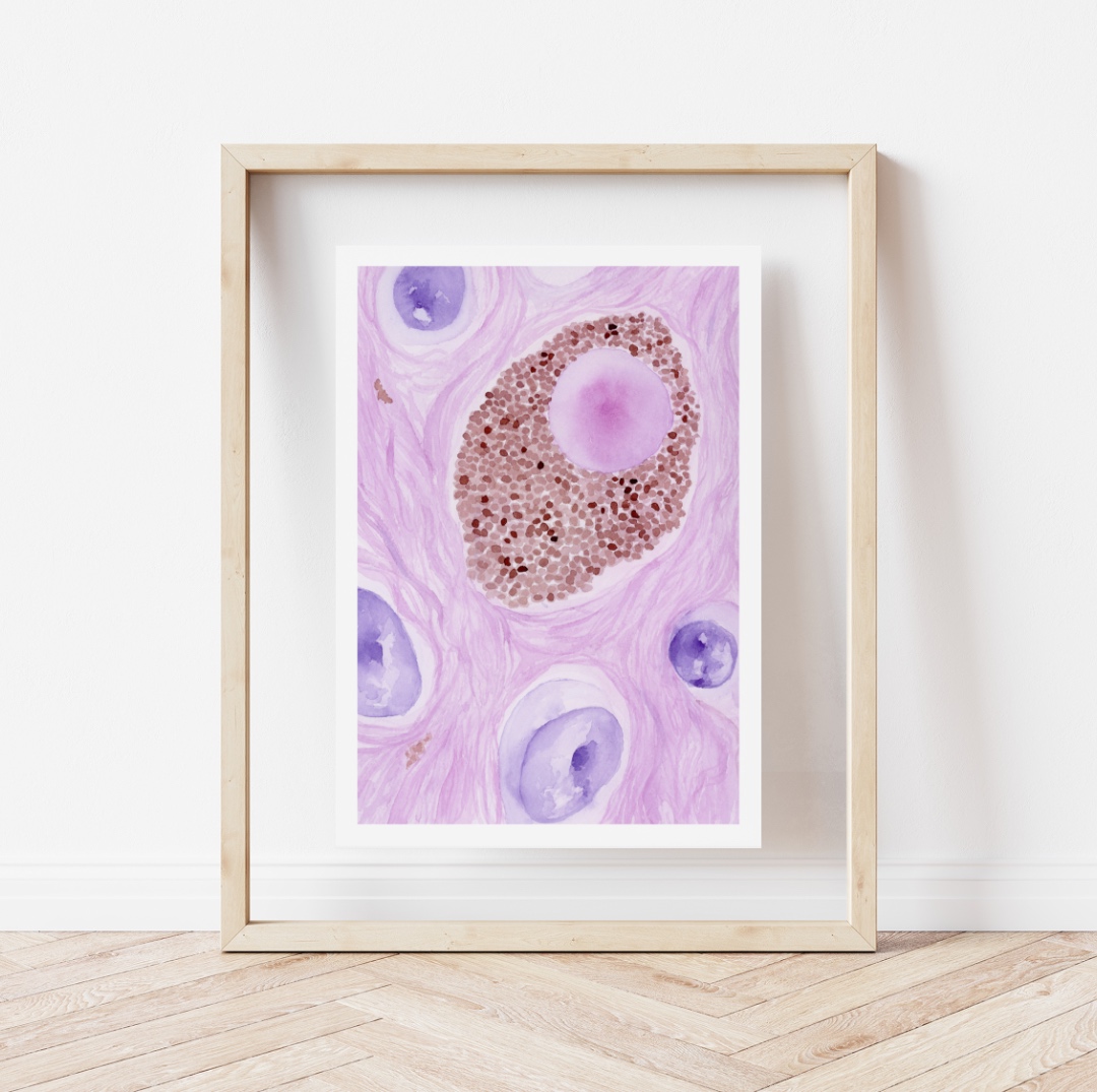
Lewy bodies are accumulations of alpha synuclein protein found in Parkinson’s disease and Lewy body dementia. They are most commonly seen in the substantia nigra in the midbrain in Parkinson’s and in the cingulate gyrus and the amygdala in Lewy body dementia. These are intracellular inclusions that can be seen on H&E (pictured) in the substantia nigra or using an alpha synuclein stain. PD and LBD can affect movement, gait, balance, memory, and cognition.
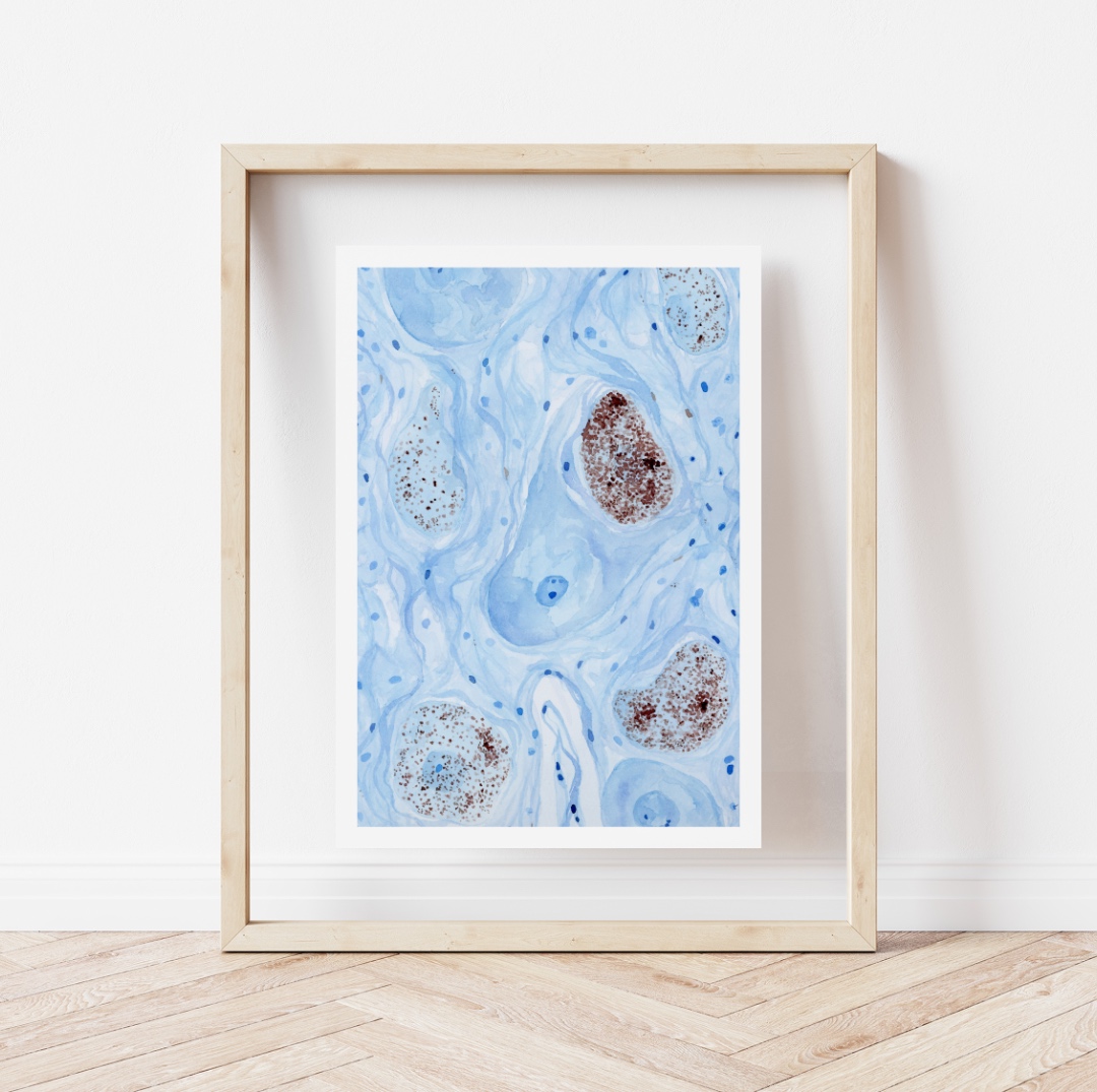
This painting is created after an image of Beta-amyloid plaques which are the other hallmark pathology of Alzheimer’s disease. These are extracellular accumulations of beta-amyloid protein that occur between neurons. Beta-amyloid pathology begins in the cortex and spreads to the brainstem with increasing disease severity. This is called the Thal phase, which is one of three scales used to determine the severity of AD. Beta-amyloid accumulation can also occur in the walls of vessels in cerebral amyloid angiopathy
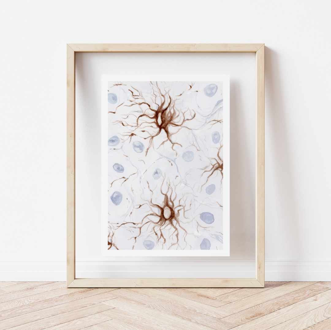
Tufted astrocytes are accumulations of tau protein near the cell body of astrocytes. These are seen in progressive supra nuclear palsy, which is a type of frontotemporal lobar degeneration (FTLD-tau). Pathology in PSP is typically seen in the motor cortex, basal ganglia, and brainstem, specifically the midbrain. The disorder causes problems with walking, balance, eye movements, and can affect speech and cognitive function in later stages.
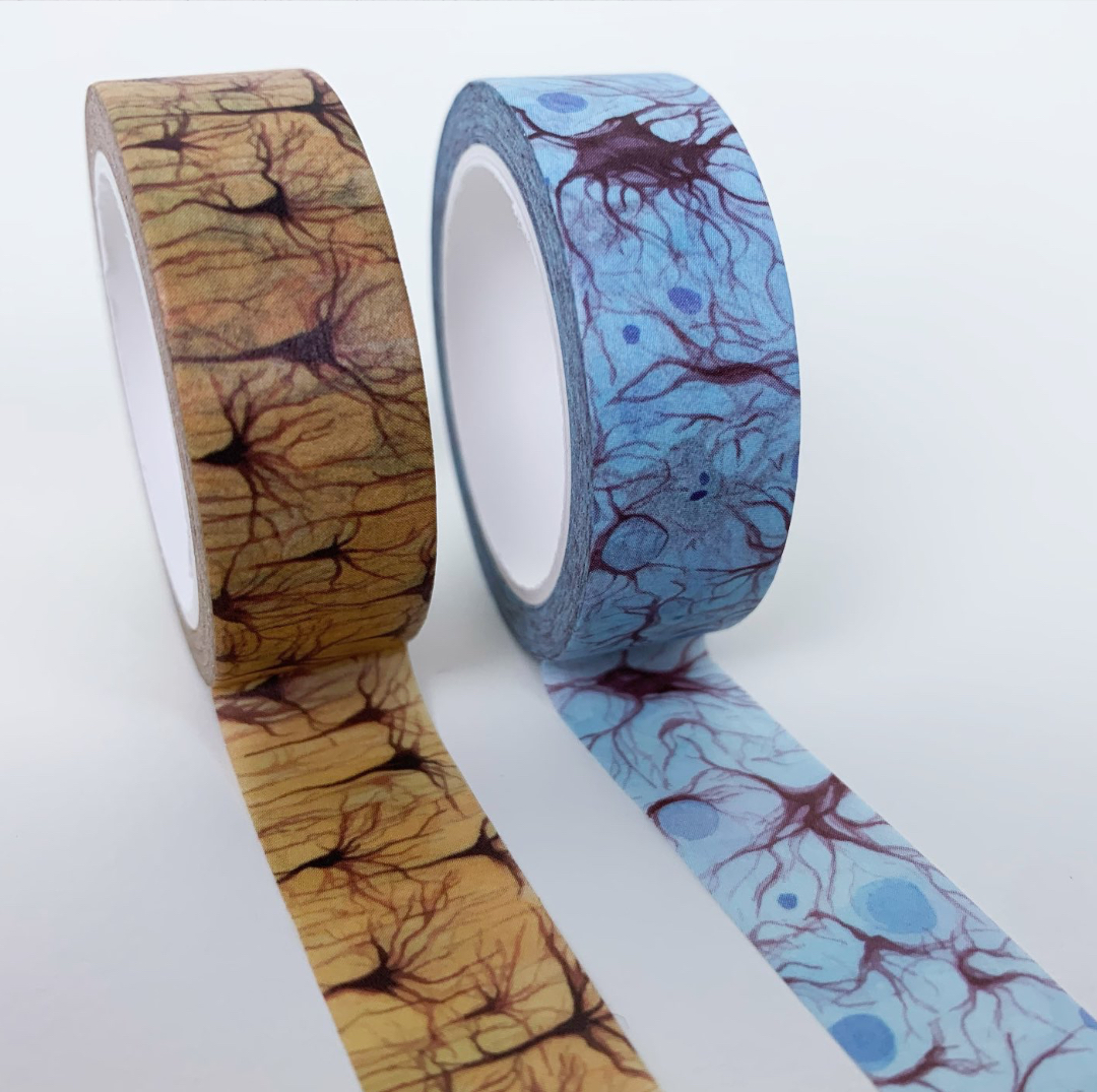
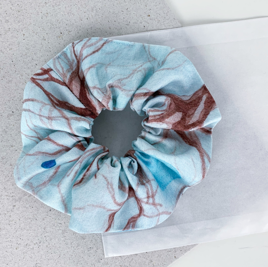
This time the shop update includes new prints & postcards from the series. Inspired by working on those brain-based paintings I created two new washi tapes with a repeating pattern showing astrocytes and neurons. And there is also a small number of neuropathology scrunchies in the shop.
